Ophthalmic Surgical Microscopes / Ophthalmic Operating Microscopes
- Home
- General Ophthalmology
- Surgical Microscope
- Ophthalmic Surgical Microscope
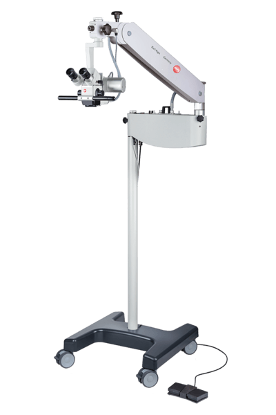
Ophthalmic Surgical Microscopes / Ophthalmic Operating Microscopes
Ophthalmic surgery needs to be very precise to handle delicate eye tissue carefully and use instruments accurately. The Ophthalmic surgical microscope / Ophthalmic Operating Microscope is one of the key tools in eye surgery. It helps eye surgeons see tiny details clearly, allowing them to work with great precision and efficiency.
An ophthalmic surgical microscope / Ophthalmic Operating Microscope is an essential tool in modern eye surgeries, including cataract surgeries, anterior and posterior segment surgeries. These microscopes are equipped with advanced features that enhance surgical precision and outcomes. One critical feature is the red reflex, which provides a clear view of the eye’s internal structures, crucial for procedures like cataract removal.
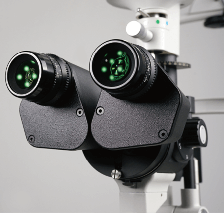
Karl Kaps ophthalmic operating microscope are renowned for their superior image quality and illumination systems. They offer wide-angle viewing systems that allow surgeons to see more of the eye at once, improving the efficiency and safety of surgical procedures. The high-quality optics ensure exceptional depth of field, enabling detailed visualization of microstructures within the eye.
The microscopes also feature adjustable light intensity to protect the retina from excessive exposure while maintaining optimal visibility. The illumination system is designed to provide consistent, bright lighting across the entire field of view, which is essential for maintaining focus and clarity during complex surgeries.
These advanced systems are vital for various segment surgeries, providing surgeons with the necessary tools to perform delicate operations with high precision. The integration of cutting-edge technology in these microscopes continues to improve surgical outcomes and patient safety.
Lab Medica Systems provides advanced surgical microscopes. These tools help ophthalmic surgeons get the best results for their patients. We supply the Karl Kaps SOM 62 Ophthal, an ophthalmic surgical microscope, in India.
Key Features of Karl Kaps SOM 62 series
- Compact and Ergonomic Design: Ideal for limited-space operating rooms, laser centers, and outpatient clinics; its modular build supports tailored expansions.
- Superior Apochromatic Optics: Provides sharp, high-contrast images with outstanding color accuracy.
- Powerful Illumination: Equipped with a 15 V/150 W coaxial cold-light illumination system featuring an integrated UV filter and retractable GG 475 retina protection for bright red reflex and enhanced patient safety.
- Effortless Positioning: Minimal manual effort required with user-friendly, intuitive controls for easy adjustments.
- Continuous Operation: Optional backup lamp and tilt-switch ensure uninterrupted surgical procedures.
- Documentation Flexibility: Supports diverse documentation needs with various stereo observer tubes, beam splitters, and adapters.
- Sterility Assured: Compatible with asepsis caps and drapes for secure sterile handling.
Ophthalmic Surgical Microscope / Ophthalmic Microscope | Karl Kaps SOM 62 series Ophthal
The surgical / operating microscopes of the SOM® 62 series are extremely space-saving and ideally equipped for surgical procedures on the eye – whether routine procedures or complex surgeries on the posterior eye.
Kaps relies on its proven modular system and offer almost unlimited equipment options, individually tailored to your requirement profile.
Applications of Surgical Microscopes / Ophthalmic Operating Microscopes
Ophthalmic Surgical Microscopes / Ophthalmic Operating Microscopes are essential tools for revealing the finest anatomical details and assisting with precise surgical maneuvers during both anterior and posterior segment surgeries. These microscopes have a broad range of applications in ophthalmology, spanning from diagnostics to intricate surgical procedures.
Anterior Segment Surgery
- Glaucoma Surgery
- Corneal Surgery
- Laser Eye Surgery
- Refractive Surgery
- Cataract Surgery
Posterior Segment Surgery
- Vitrectomy
- Retinal Detachment Repair
- Macular Hole Repair
- Maculopathy Surgery
- Posterior Sclerotomy
- Radial Optic Neurotomy
- Macular Translocation Surgery
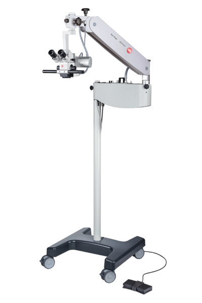
SOM© 62 Ophthal Basic
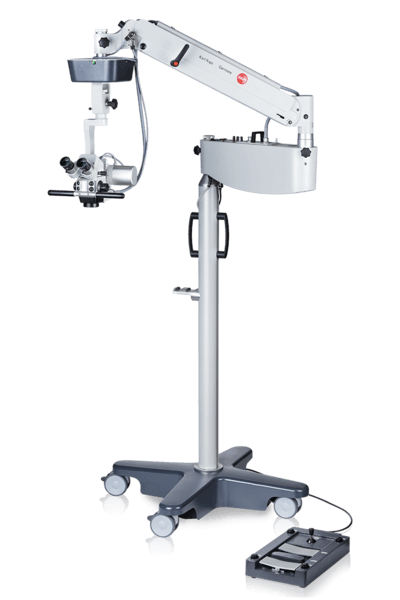
SOM© 62 Ophthal Basic +
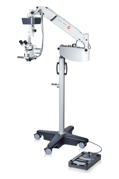
SOM© 62 Ophthal Advanced
Ophthalmic Surgical Microscope / Ophthalmic Operating Microscope Technical Specification
| SOM© 62 Ophthal Basic | SOM© 62 Ophthal Basic + | SOM© 62 Ophthal Advanced | |
| Binocular tube | Inclined tube 60°, f = 159 mm | Inclined tube 60°, f = 159 mm | Swivel tube 0 – 210°, f = 182 mm |
| Objective | APO f = 200 mm | APO f = 200 mm | APO f = 200 mm |
| Eyepieces | Eyepieces WF 10 x V with diopter compensation, Eyepieces WF 12.5 x V with diopter compensation (optional), Other on request | ||
| Magnification | Apochromatic 5-step magnification changer | Apochromatic 5-step magnification changer | Apochromatic motorized zoom 1:6 |
| Fine focusing | 40 mm motorized | 40 mm motorized | 40 mm motorized |
| XY-coupling | – | 50 x 50 mm | 50 x 50 mm |
| Filter | Swing in retina protection filter GG 475 Swing in yellow filter | ||
| Eyepieces | Coaxial cold light fiber optic illumination 15 V / 150 Watt with operational replacement lamp, retina protection filter GG 475, filter against IR and UV exposure | ||
| Accessories | SOM© 62 Ophthal | ||
| Photo-/video documentation | Beam splitter monolateral or bilateral Beam splitter with c-mount connection, f = 54 mm TV-tube with aperture f = 54 mm Ikegami HD video camera (medical grade) |
||
| Second observer attachments | Binocular assistant tube Binocular inclined tube, f = 125 mm / swivel tube 0 – 210°, f = 182 mm IEyepieces WF 10 x V with diopter compensation |
||
| Others | Monitor holder on swivel arm Monitor 22″ (medical grade) |
||
Ophthalmic Surgical Microscope
What Are Surgical Microscopes?
A surgical microscope, also known as an operating microscope, is a specialized optical microscope designed for use in surgical procedures, particularly microsurgery.
What to Expect From Surgical Microscopy?
A surgical operating microscope helps surgeons see a patient’s anatomy in high detail. It offers clear visibility of small details with great resolution and contrast.
These microscopes improve the surgical view with special technologies. They use augmented reality fluorescence for neurovascular procedures. They also have built-in OCT for advanced eye surgeries.
Compatibility with devices like IGS systems, endoscopes, and DICOM enhances the surgical view. This helps surgeons focus on the patient and supports their decision-making.
What Are the Key Factors to Consider When Selecting a Surgical Operating Microscope?
When selecting a surgical microscope, consider the following factors: optical quality, low-light illumination design for enhanced patient safety, ergonomic design with adjustable binoculars, workflow support, device connectivity within the OR, and hygiene considerations.
What is a Surgical Microscope Used for?
Surgical microscopes provide sharp, clear images, flexibility, ease of use, and high-quality video and recording systems. Surgeons in neurology, ophthalmology, ENT, and plastic reconstructive surgery rely on these microscopes to magnify the surgical site, revealing details beyond what the naked eye can see.
Recent technological innovations have greatly improved surgical microscopes, enhancing performance and expanding diagnostic and operative capabilities.
Notable advancements include smaller horizontal optics and better reach for easier movement. There are ergonomic designs for surgeon comfort.
The system has fully integrated HD video in both 2D and 3D. It also features intraoperative fluorescence to see blood flow or tumors. Lastly, it provides a stable red reflex for precision. These developments have ultimately improved surgical outcomes and patient care.
Why Use a Surgical Microscope?
For delicate surgical procedures, visualization solutions are crucial, enabling surgeons to operate under optimal conditions.
Increasingly, surgeons are opting for microscopes over binocular glasses due to the significantly higher magnification, allowing for the visualization of the finest details.
Microscopes offer additional advantages, including extended depth of field, high-resolution imaging, freedom of movement, ergonomic posture, and integrated visualization technologies with augmented reality, providing greater confidence in surgical decision-making.
Frequently Asked Questions Surgical Microscopes
Lab Medica System provides an extensive selection of surgical operating microscopes designed for both diagnostic applications and a broad range of clinical microsurgical procedures. These microscopes support surgeries across various fields, including anterior and posterior segment ophthalmology, neurosurgery (spinal and cranial), ENT surgery, plastic and reconstructive surgery, and dental surgery.
A surgical operating microscope enables surgeons to observe a patient’s anatomy with high magnification, allowing for clear visualization of the finest details with excellent resolution and contrast. These microscopes also improve surgical precision through integrated visualization technologies, such as augmented reality fluorescence for neurovascular procedures and built-in OCT for complex ophthalmic surgeries. Additionally, compatibility with devices like IGS systems, endoscopes, and DICOM enhances the surgical view, aiding surgeons in maintaining focus on the patient and facilitating informed decision-making.
In delicate surgical procedures, advanced visualization solutions are crucial for ensuring optimal conditions during surgery. Increasingly, surgeons are choosing microscopes over binocular loupes due to the superior magnification that allows for detailed visualization.
Beyond magnification, surgical microscopes offer additional advantages, including an extended depth of field, high-resolution imaging, enhanced freedom of movement, ergonomic posture, and integrated visualization technologies such as augmented reality. These features provide greater confidence in surgical decision-making and overall precision.
At Lab Medica System, ongoing, long-term support is a core priority. Our commitment begins with production and extends through assembly, shipment, delivery, installation, and the maintenance programs we provide. We understand that in today’s world, offering products alone is not enough.
Authorization Letter
