Mediworks Digital Slit Lamp At Best Price in India
Mediworks Digital Slit Lamp
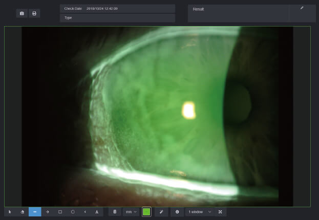
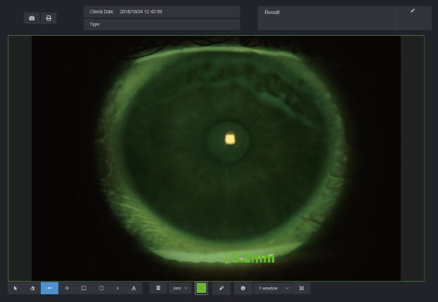
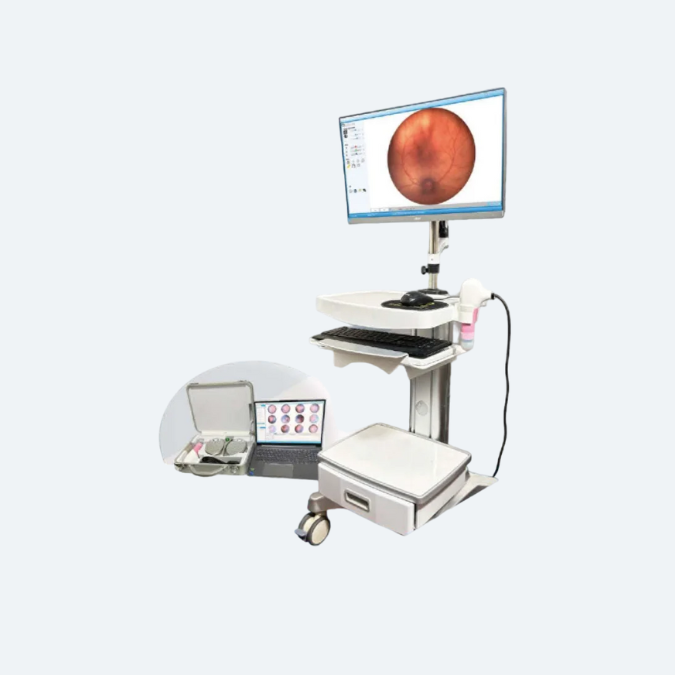
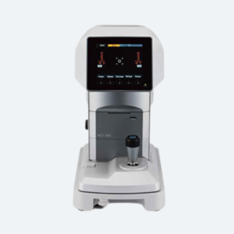
Digital slit lamp is a widely used device used for an eye exam. With the help of this tool, ophthalmologist can get a closer look of the front and the inside of the eye. The simple and compact designed slit lamp is best when it comes to saving space and offers easy operations. We are Importer, Wholesaler & Supplier of Mediworks S390L Slit Lamp in India.
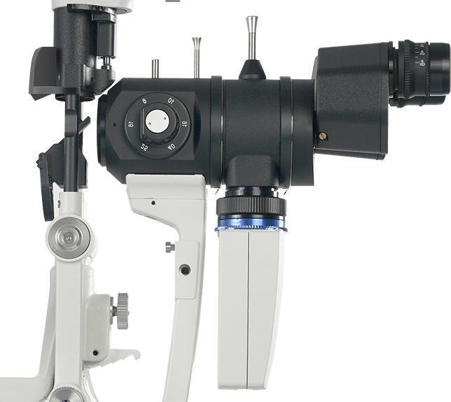
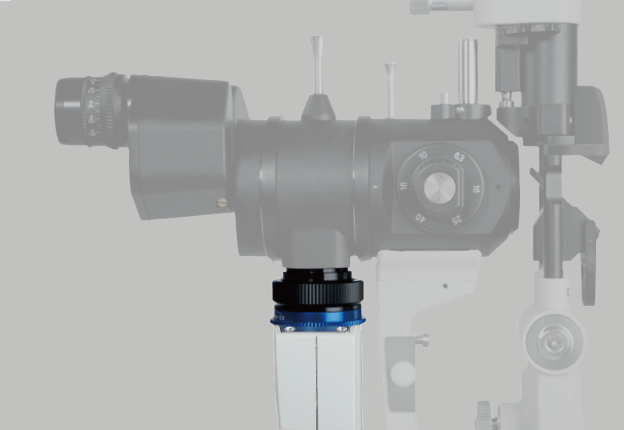
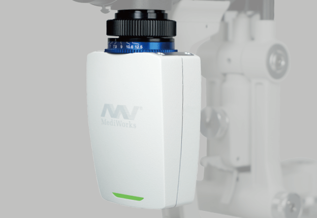
The design of the ophthalmic slit lamp S390L(Firefly WDR) was inspired by the shape of the firefly. The smart design largely saves space for clinicians compared to other bulky camera systems. We have preset many camera parameters so the user does not need to adjust settings before using the device. The user can operate the machine immediately once the installation has been finished. The device has the following automatic functions for photo shooting and processing when equipped with our Mediview software:
Wide Dynamic Range
Meibomian Glands Examination
Auto Exposure
Auto Gain
Auto White Balance
Auto OS/OD Indicator
High sensitivity. The slit is still clear and sharp under weak light.
Wide dynamic range. Iris and sclera images are simultaneously clearly presented with more realistic and evenly distributed color
HD Optical System
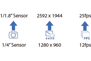
Optical resolution is up to 200 lp/mm(2700·N lp/mm), providing more details of the pathologies.
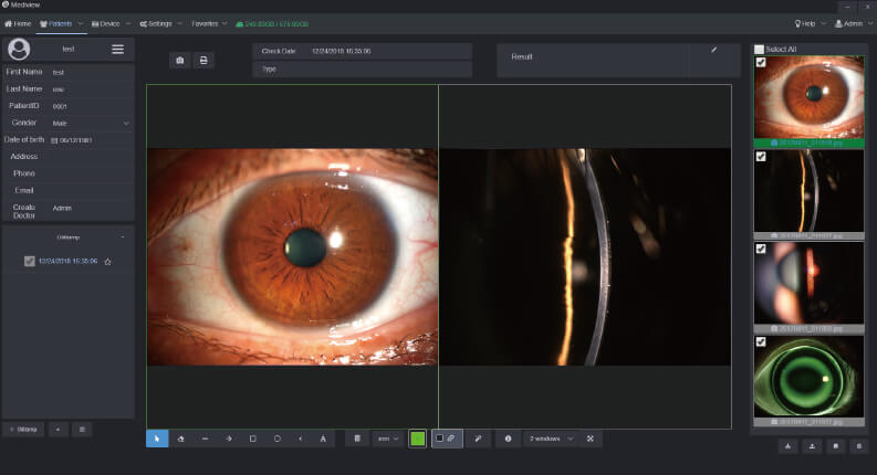
Built-in yellow filter
The built-in yellow filter of the ophthalmic slit lamp microscope helps doctors to get a clear tissue image with enhanced contrast.
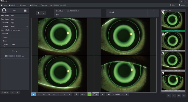
The Meibomian glands examination
The unique infrared light source module supports the observation of the patient’s Meibomian gland.
Software Features
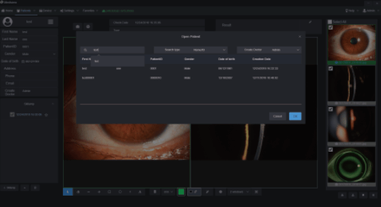
Convenient Patient Management
The patient management system enables clinicians to build and edit patient record,search information by inputting keywords.
Clinicians can easily record symptoms and manage the data all the time.
The software supports DICOM which makes the images captured by Firefly be easily integrated into hospital’s medical system.
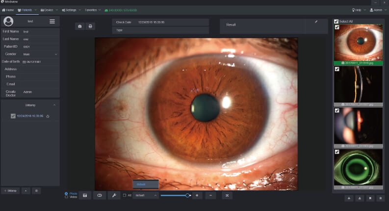
Functional Image Analysis
Clinicians can measure the pathology area with our powerful software tools and change the contrast and brightness of the images. Clinicians can also compare several images at one time to analyze the symptoms and pathology.
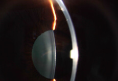
Orthokeratology Lens Fitting Assistance
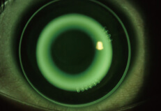
The optometrists can capture and record high resolution fluorescein images of lens fitting and real-time video without a recording time limit. By comparing the different lens fitting effects, the optometrist can show and educate patients which lens is most suitable for them.
Customized Auto Exposure Value Setting
Clinicians can customize auto exposure values according to the image demand and save as templates for future capturing purpose.
Also, the printing report can be customized according to clinician’s needs.
The Most Effective Tools for Dry Eye
Meibomian Gland Observation
Built-in infrared light source allows the doctor to accurately judge the absence of the meibomian glands.
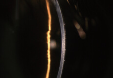
Tear Film Break-up Time
High-performance digital module, doctors can get the tear film break-up time and judge the stability of it by high-resolution video recording.
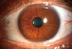
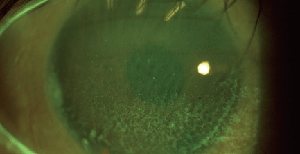
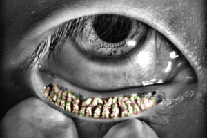
Red Eyes Analysis and Keratopathy Exposure
With a built-in yellow filter, doctors can accurately analyzemeye surface damage and inflammation images.
Tear Meniscus Height
Doctors can obtain tear meniscus height by using measuring function in the Mediview software, and effectively evaluate tear meniscus height.