Dry Eye Diagnostic System​

Diagnostic System of Dry Eye

We are Importer, Wholesaler & Supplier of Mediworks D130 Dry Eye Diagnostic System in India.


Dry eye is a multifactorial disease of the tears and ocular surface that results in symptoms of discomfort, visual disturbance, and tear film instability with potential damage to the ocular surface.


Mediworks D130 system enhances accurate diagnoses and earlier intervention, providing guidance for customized treatment.


Dry Eye Diagnostic System can provide a standardized examination and quantified causes for the evaluation of Dry Eye Disease.


The system uses various techniques such as Schirmer’s test, Tear Film Osmolarity test, Meibomian Gland Imaging, and Lipid Layer Thickness measurement to diagnose Dry Eye Disease.


The diagnosis of dry eye disease is based on a combination of symptoms, clinical signs, and diagnostic tests.

Diagnostic System of Dry Eye, Mediworks D130

Dry Eye Diagnostic System

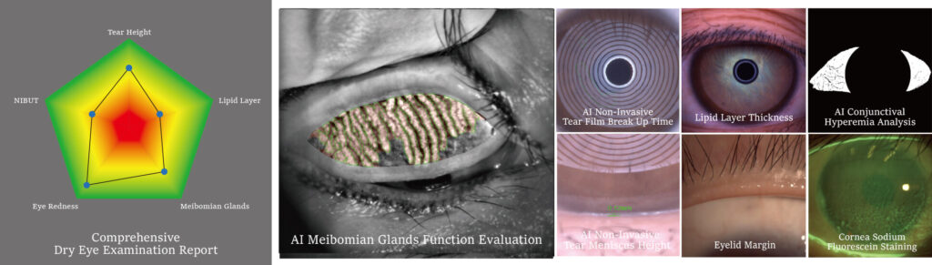
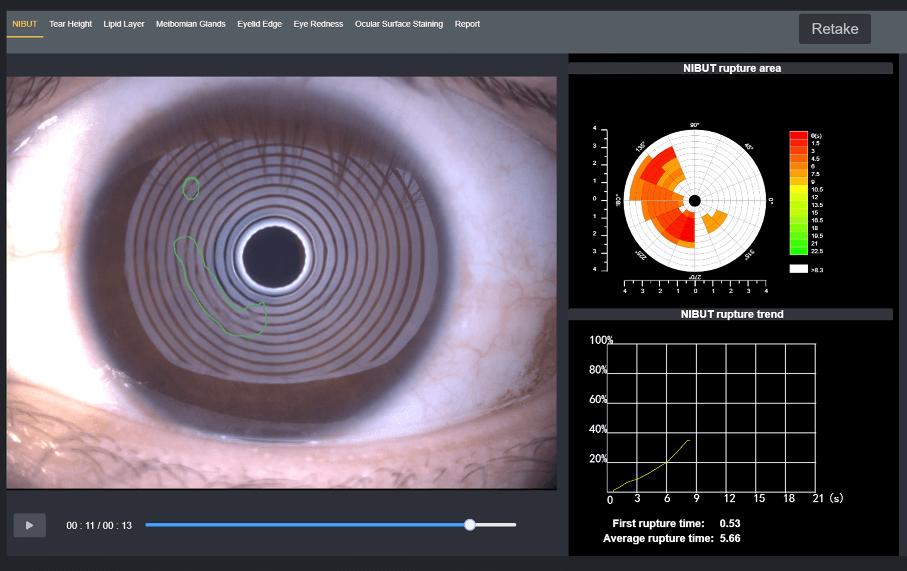
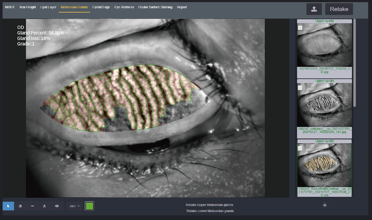
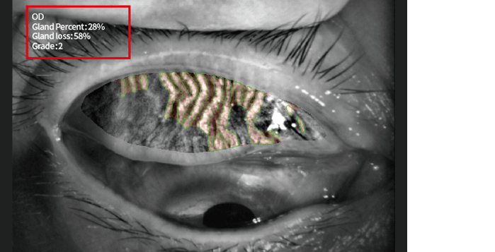
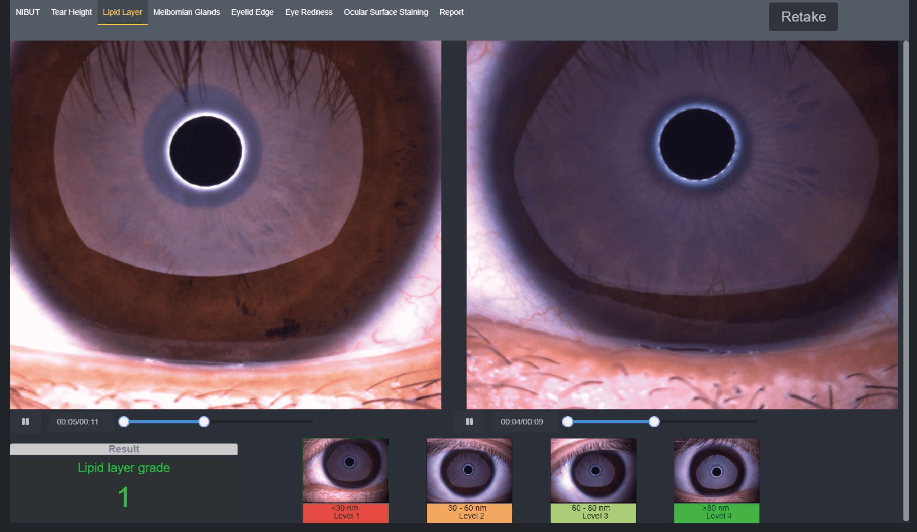
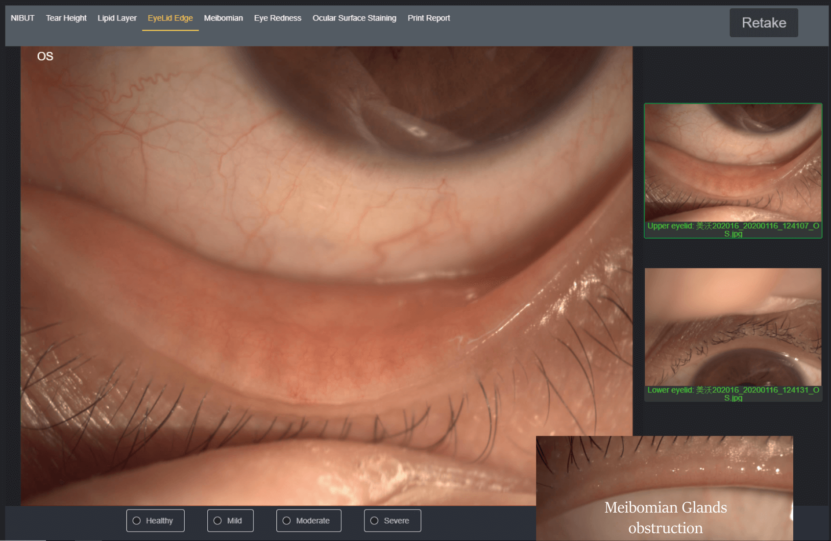
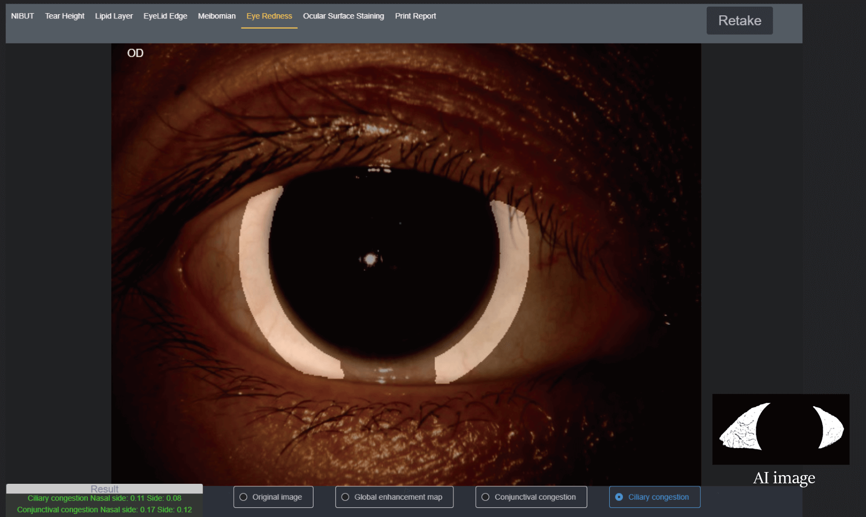
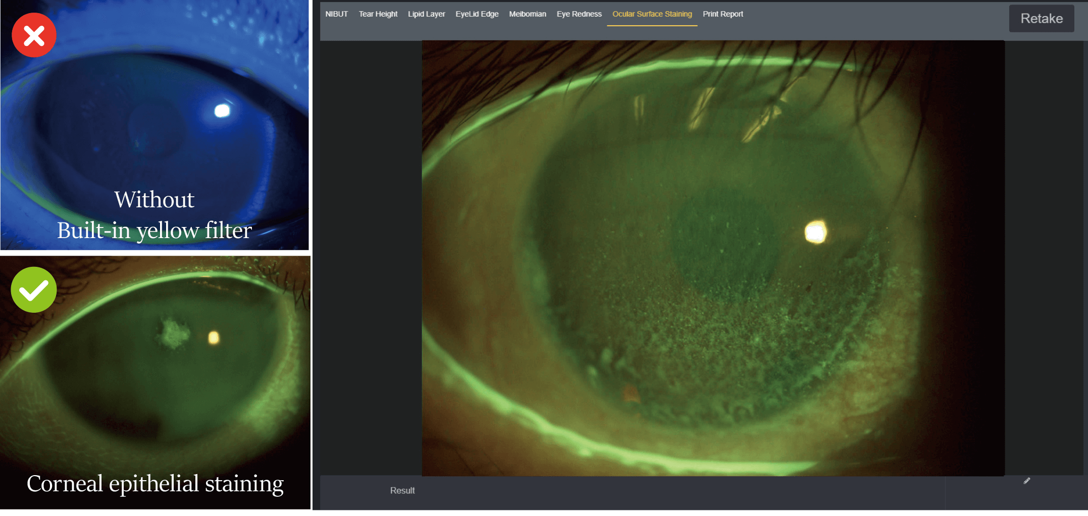
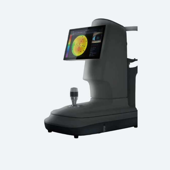
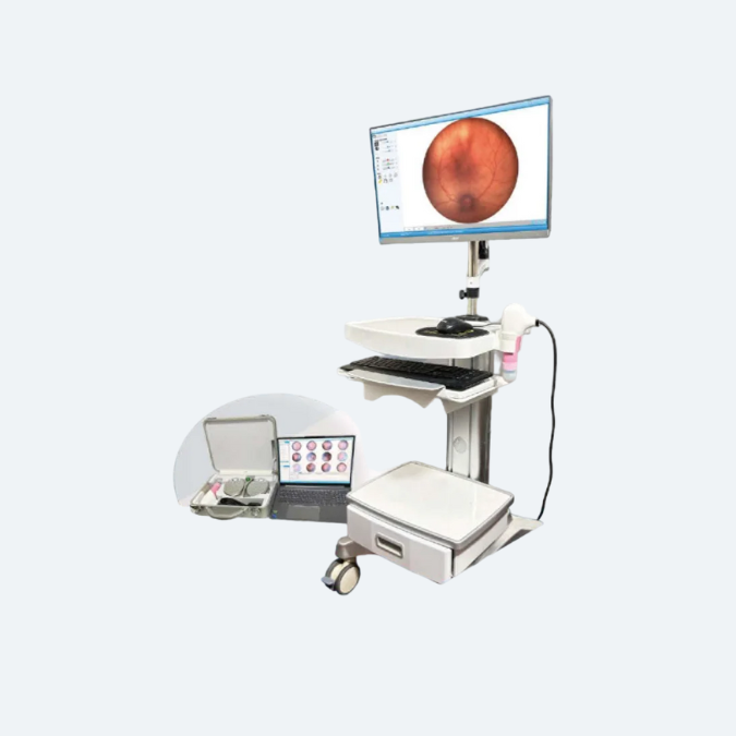
Dry Eye Diagnosis / AI Meibomian Glands Analysis


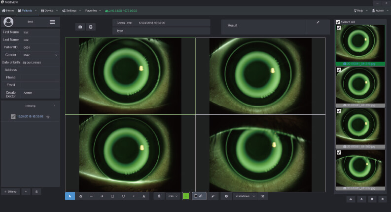
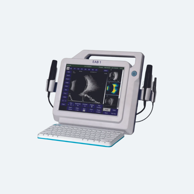
Anterior Segment Photography / Lens Fitting
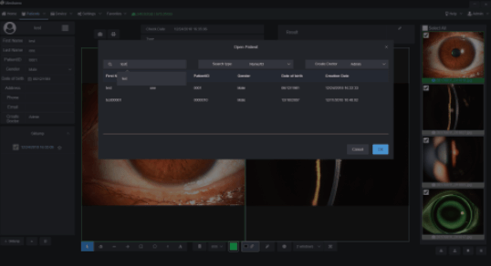
 

Patient Management


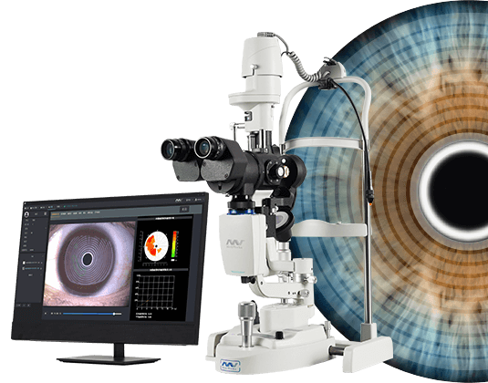
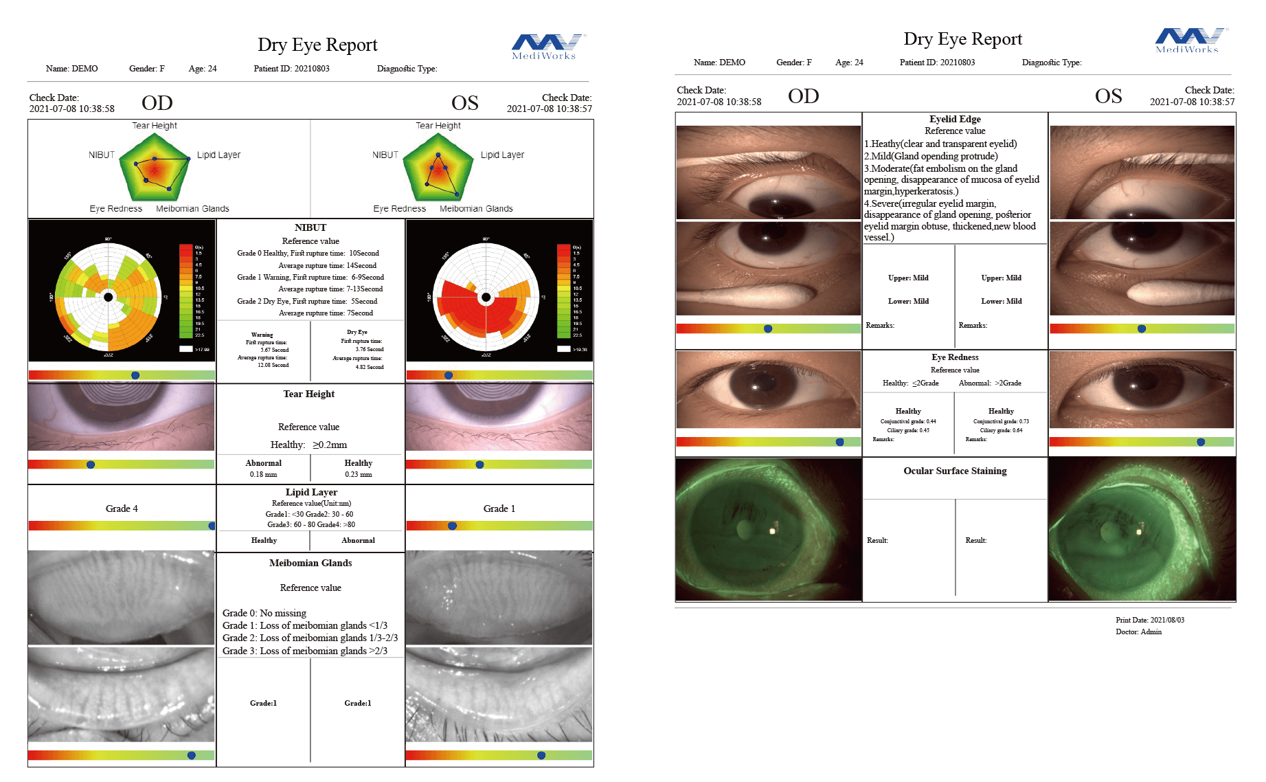
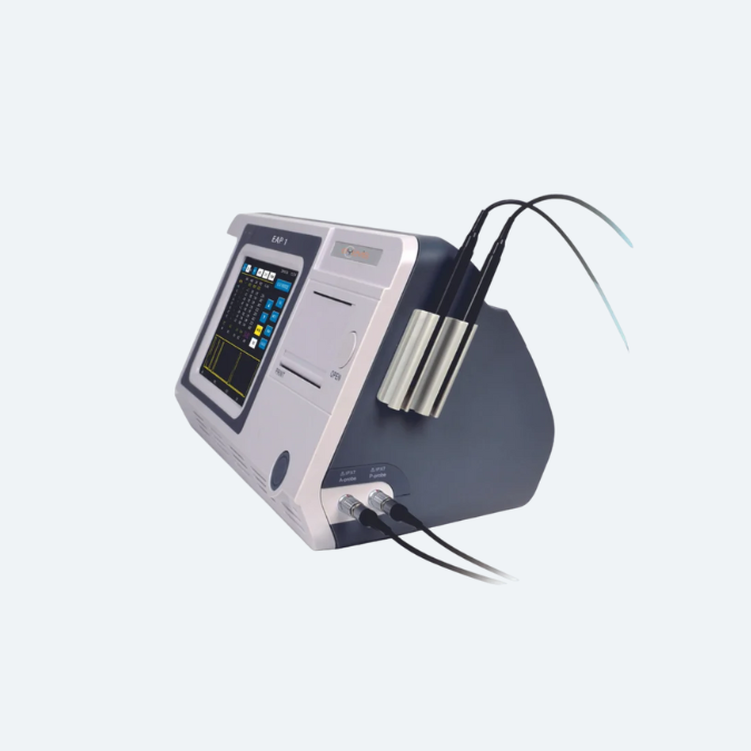
As an excellent dry eye device, our dry eye diagnostic system enhances accurate diagnoses and earlier intervention, providing guidance for customized treatment.

Platform for Comprehensive Ocular Surface Examination

Dry eye diagnosis/Anterior Segment Photography/Lens fitting/ Patient management/Telemedicine


Guided examination:providing a comprehensive report covering 7 dry eye diagnosis.Non-invasive examination,Quantitative data.


Full-automatic Firefly digital module ,easy operation without parameter settings.High quality optics and built-in yellow filter efficiently increase the accuracy of lens fitting.


Professional 1/1.8-inch sensor and 2.4μm pixel,real-time playing and storage. Smart patient management system,DICOM supported.

Due to various causes of Dry Eye Disease,traditional examination is difficult to find out the cause and quantify for the diagnosis. MediWorks Dry Eye Diagnostic System can provide standardized examination and quantified causes evaluation for Dry Eye Disease.

Overview

AI Non-Invasive Break Up Time

After taking one video, it brings out automatis result of NIBUT and Tear Meniscus Height.

AI identifies the break-up area and analyzes NIBUT automatically. Fully automatic analysis system provides efficient quantified evaluation for the overall stability of tear film.It automatically acquires the first break up time, average break up time, break up distribution,break up area percentage curve and time distribution.


  • Grade 0 Normal, First Rupture Time: 10 s Average Rupture Time: 14 s
  • Grade 1 Warning, First Rupture Time: 6-9 s Average Rupture Time: 7-13 s
  • Grade 2 Dry eye, First Rupture Time: 5 s Average Rupture Time: 7 s

MediWorks adopts Placido ring projection system with visible light to do NIBUT examination,the examination scope is up to 8mm cornea diameter which brings much more comprehensive diagnosis outcome.


The non-invasive examination avoids the irritation brought by the traditional Cornea Sodium Fluorescein Staining.

AI Non-Invasive Tear Meniscus Height

  • Normal: ≥0.2mm

AI identification system depicts Tear Meniscus area and measures the tear height automatically. Evaluate tear secretion amount and continuity objectively.More efficient and less irritation compared with the traditional Schirmer’s test.

  • Insufficient tear secretion
  • Abnormal dynamics and conjunctival chalasis

Evaluation of Meibomian Glands Function

Get original/enhanced/result images by one click. AI identification system automatically anlalyzes meibomian glands loss. caused by meibomian glands dysfunction with precise and quantified diagnosis results.


Built-in infrared lighting system helps doctors obtain larger image scope of the meibomian glands.


Adjustable depth of field makes the glands more prominent and distinguishable against the background.


  • Grade 0: No Meibomian Glands Loss
  • Grade 1: Meibomian Glands Loss < 1/3
  • Grade 2: Meibomian Glands Loss 1/3-2/3
  • Grade 3: Meibomian Glands Loss >2/3
  • Meibomian glands loss
  • Image of Meibomian Glands under high-magnification

Lipid Layer Thickness

White ring projection system ensures a larger examination area compared to Placido ring.By comparing with the standard grading template and recording the Lipid Layer thickness, it is helpful for judging MGD.


  • Grade 1: < 30
  • Grade 2: 30-60
  • Grade 3: 60-80
  • Grade 4: >80
  • (Unit:mm)

Eyelid Margin

MediWorks professional design of optical system is capable of providing HD digital image that remains clear and sharp even zoom in, meets the examination requirements of the overall shape of eyelid margin and its slight change.


  • Normal including (Ophthalmic embolism bright, transparent)
  • Mild including (gland cap crown – glandular prominent)
  • Moderate including (glandular fat plug – disappearance of the marginal mucosa, hyperkeratosis)
  • Severe including (uneven margins, disappearance of the meibomian glands – posterior margin Blunt round, thickening, new blood)

AI Analysis of Conjunctival Hyperemia

  • Normal: ≤2 Abnormal: >2

The unique AI identification system can identify and calculate percentages of conjunctival congestion and ciliary congestions and evaluate severity of eye congestion.

Cornea Sodium Fluorescein Staining

Effectively increases positive rate of early corneal epithelial staining.

Built-in yellow filter along with cobalt-blue filter makes the corneal sodium fluorescein images more clearly.

Dry Eye Comprehensive Evaluation Report

Convenient Medical Consultation on Dry Eye Syndrome

Smart Patient Management system

Comparison of Patient records

Smart Patient Management system supports repeated comparison among medical records to help doctors develop customized treatment plans and evaluate treatments.

Patient Management system allows doctors to build and edit medical records, and quickly search the patient case by key words. Besides, doctors can note patients’ situation via the software. With the DICOM-supported system, Mediview is connected with medical systems in hospitals.

Mediworks D130 Dry Eye Diagnostic System Specification

Microscope
Microscope Type Galilean Type
Magnification Change Revolving Drum 5 steps
Total Magnification 6.3X, 10X, 16X, 25X, 40X
Optical Resolution 2700·N lp/mm (200 lp/mm)
Eyepieces 12.5X
Angle between Eyepieces 10°
Pupillary Adjustment 52mm-80mm
Diopter Adjustment -8D~+8D
Field of View Ø36.2mm, Ø22.3mm, Ø14mm, Ø8.9mm, Ø5.7mm
Slit Illumination
Slit Width 0~14mm continuous (slit becomes a circle at 14mm)
Slit Length 1~14mm continuous
Aperture Diameters Ø14mm, Ø10mm, Ø5mm, Ø3mm, Ø2mm, Ø1mm, Ø0.2mm
Slit Angle 0°~180°
Slit Inclination 5°, 10°, 15°, 20°
Filters Heat-absorbing filter, ND filter, Red-free filter, Cobalt blue filter,Yellow filter built-in
Lamp LED
Luminance ≥150klx
Power Supply
Input Voltage 100V~240V
Input Frequency 50Hz/60Hz
Rated current 1.2A
Output Voltage 3V LED, Fixation 15V
System Specifications
Digital Module Automatic exposure/ Automatic white balance / Adjustable depth of field and aperture
Image Sensor 1/1.8-inch sensor / 2.4μm pixel / 5.0M Pixels
Photo Resolution 2592 x 1944
Frame of Video 25fps
Video Formats MP4 H.264
Exposure Mode Automatic exposure
Transmission Interface USB
Packaging
Dimension 740mm x 450mm x 530mm(L/W/H)
Gross weight 23kg
Net weight 17kg
Computer Specifications
PC configuration i5-10500T 8G memory 25GB SSD+1TB storage
Display 1920×1080 23.8inch
PC system Windows 10

Dry Eye Module

AI Non-Invasive Tear Break Up Time AI Non-Invasive Tear Meniscus Height
AI identify the break-up area AI identification system
Automatic first break up time Automatic Non-Invasive Tear Meniscus Height
Automatic average break up time Optical magnification
Visible light Placido ring projection(23 ring) Electronic amplification
AI Conjunctival Hyperemia Analysis Cornea Sodium Fluorescein Staining
AI identification system Eye surface damage report
Automatic conjunctival congestion percentages Built-in yellow filter
Automatic ciliary congestions percentages Cobalt blue filter
AI Meibomian Glands Function Evaluation Lipid Layer Thickness
AI identify Meibomian glands Template comparison evaluation
Automatic Meibomian glands loss classification Visible light White ring projection system
Eyelid Margin Dry Eye Examination Report
Optical magnification Automatic analysis report
Electronic amplification

Reach Out