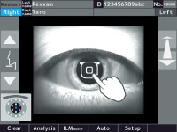
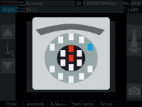
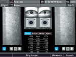
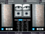
| Capturing of corneal endothelial cell | Capturing position | Capturing range | 0.25mm×0.55mm(W×H) |
| Center | 1 point |
| Paracenter | 6 points(2,4,6,8,10 and 12 o’clock directions) |
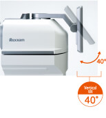
| Periphery(optic angle:27 degrees) | 10 points(1,2,4,5,6,7,8,10,11 and 12 o’clock directions) |
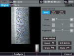
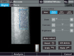
| Measurement of corneal thickness | Range of corneal thickness measurement | 400 to 750μm(step:1μm) |
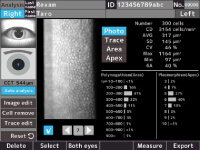
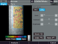
| Analysis parameter | [Number] [cells] | Number of endothelial cells |
| [CD] [cells/m㎡] | Density of endothelial cells |
| [AVG] [μ㎡] | Average endothelial cell area |
| [SD] [μ㎡] | Standard deviation of cell area |
| [CV] [%] | Coefficient of variation of cell area |
| [Max] [μ㎡] | Max.cell area |
| [Min] [μ㎡] | Min.cell area |
| [6A] [%] | Rate of cell hexagonality |
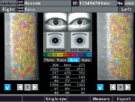
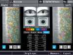
| Histogram | Polymegathism |
| Pleomorphism |
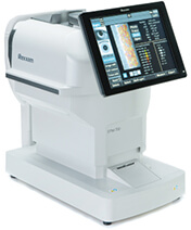
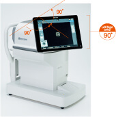
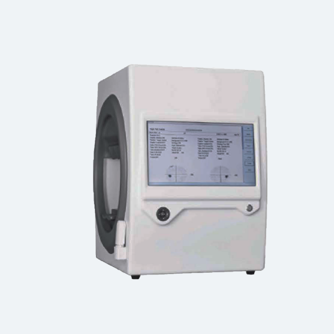
| Monitor | 10.4 inch touch panel colored LCD(XGA) |
| Printer | Thermal line printer (paper width 58mm) |
| External interface | Ethernet(10/100Mbps)×1、USB-A×2、USB-B×1 |
| Source voltage/frequency | AC100V – 240V、50/60Hz |
| Power consumption | 90VA |
| Power saving function | OFF,3,5,10min. (switchable) |
| Size | H(503mm)×W(271mm)×D(459mm) |
| Weight | 19kg |
| Standard Accessories | Operation manual, Power cord, Printer paper, Fuse, Dust cover, Chinrest paper, Chinrest paper pin |